Smile transformations in Norwich
Discover real patient transformations from Invisalign, composite bonding, dental implants and cosmetic dentistry - trusted by Norwich patients for over 30 years.

Discover real patient transformations from Invisalign, composite bonding, dental implants and cosmetic dentistry - trusted by Norwich patients for over 30 years.
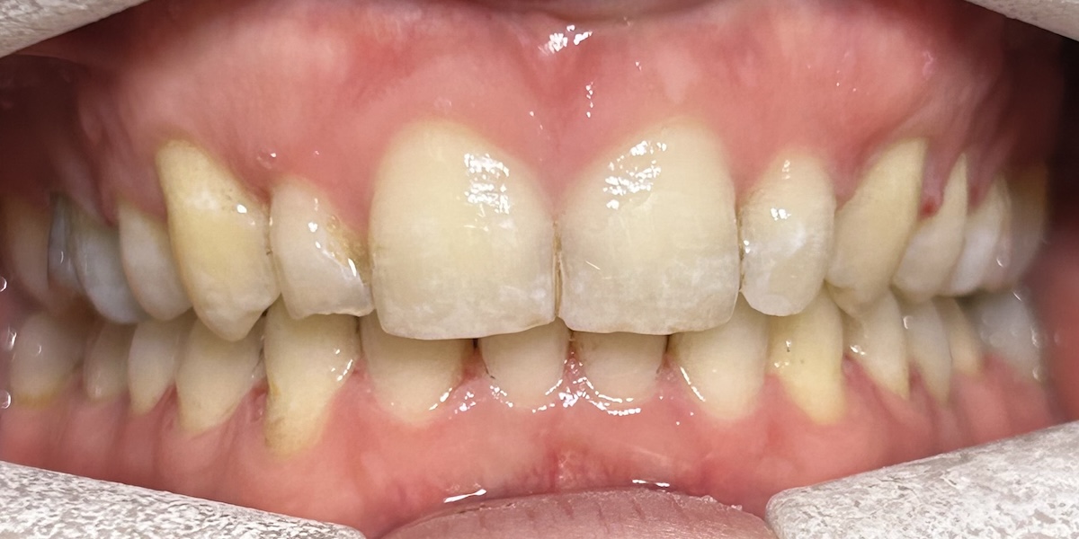
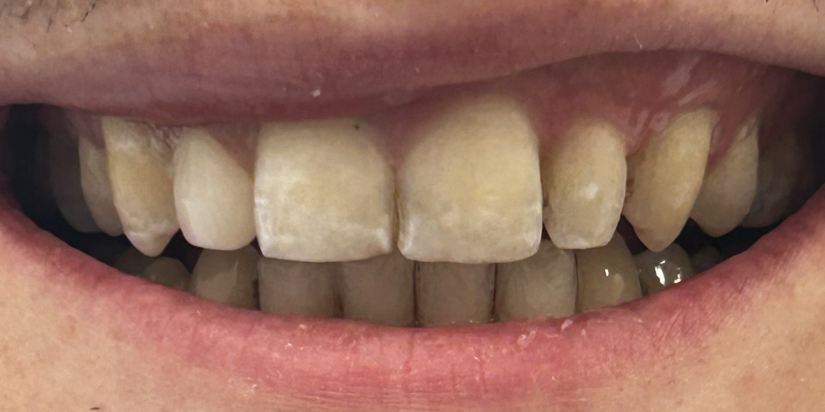
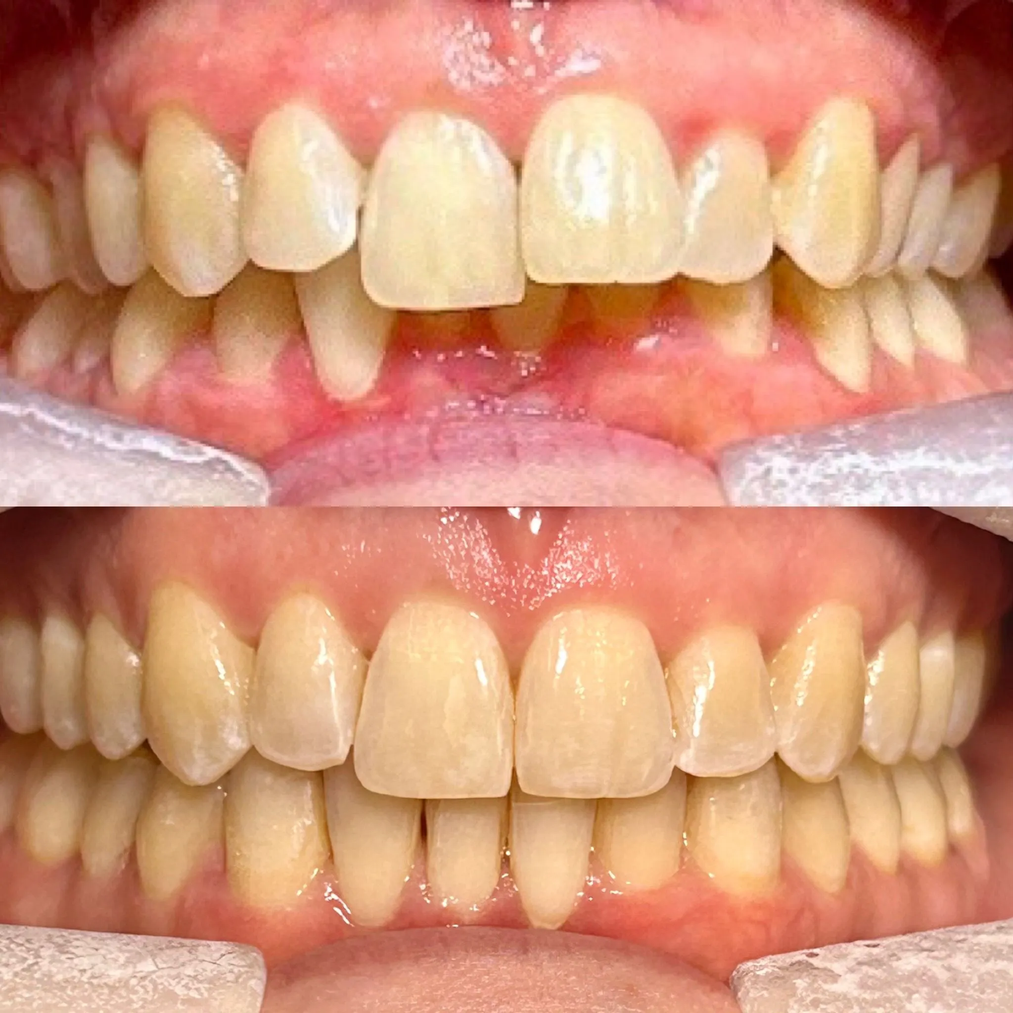
The patient self-referred for an orthodontic assessment, presenting with the chief complaint that her teeth are not straight. She expressed a specific interest in treatment with fixed orthodontic braces.
The patient’s medical history was checked verbally and revealed no contraindications to treatment.
On clinical examination, there is evidence of palatal erosion affecting UR2 and tooth surface loss affecting the lower anterior teeth. Both UR2 and UL2 are noted to be small and underdeveloped. Oral hygiene is assessed as good to average, and the patient reports no current oral habits.
Skeletal assessment reveals a Class II anteroposterior relationship, with overall facial symmetry and a low to average Frankfort mandibular plane angle (FMPA). The incisor relationship is Class II Division 2, with an overjet of 3.5 mm and an increased, complete overbite.
Dental midlines are discrepant, with the upper midline deviated to the right and the lower midline deviated to the left, resulting in a 2 mm discrepancy between arches.
Molar relationships are Class II half-unit bilaterally. Canine relationships are asymmetric, with the right canine in Class I and the left canine in Class II half-unit. A scissor bite is present involving UL5. No spacing is present in either arch; however, moderate crowding is evident in both the upper and lower arches.
Based on the clinical findings, the case was assessed as IOTN grade 4D, indicating a definite need for orthodontic treatment.
As part of the diagnostic work-up, upper and lower impressions were taken and sent to Nova Laboratory.
Comprehensive intraoral and extraoral photographs were obtained, and an OPG radiograph was taken at today’s visit. The patient was informed that a treatment plan will be formulated and discussed at the next appointment once all records have been reviewed.
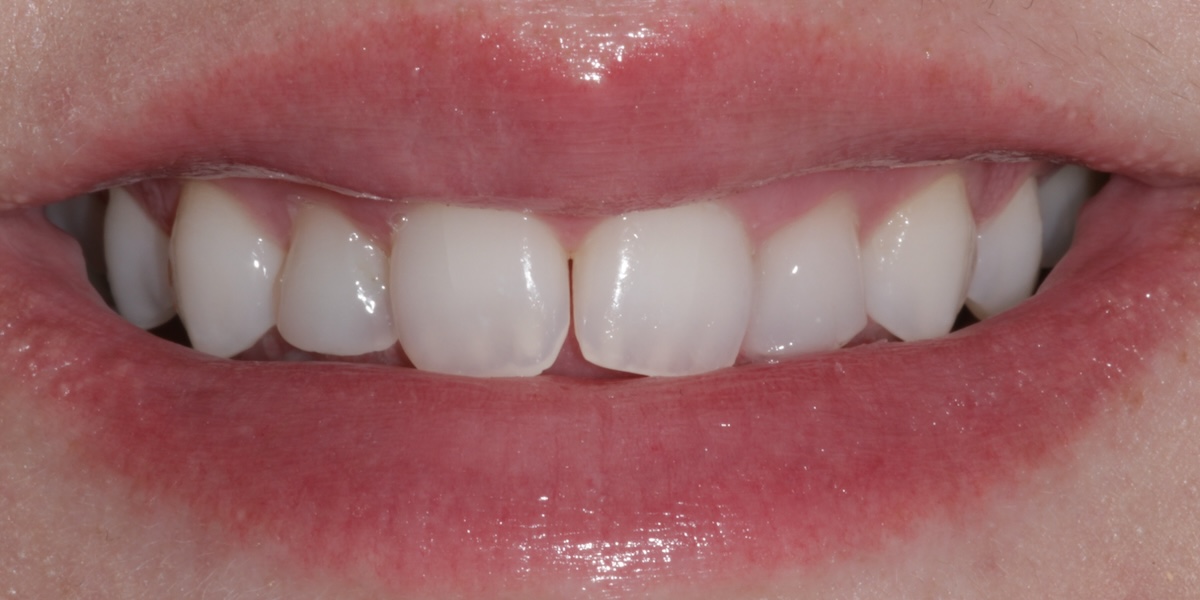
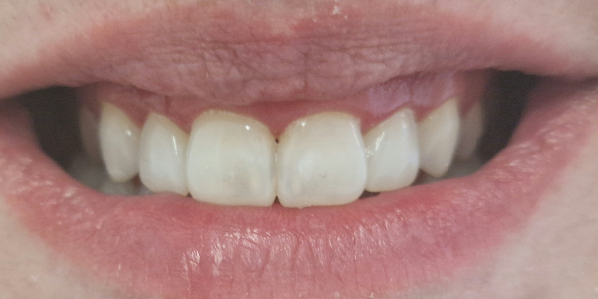
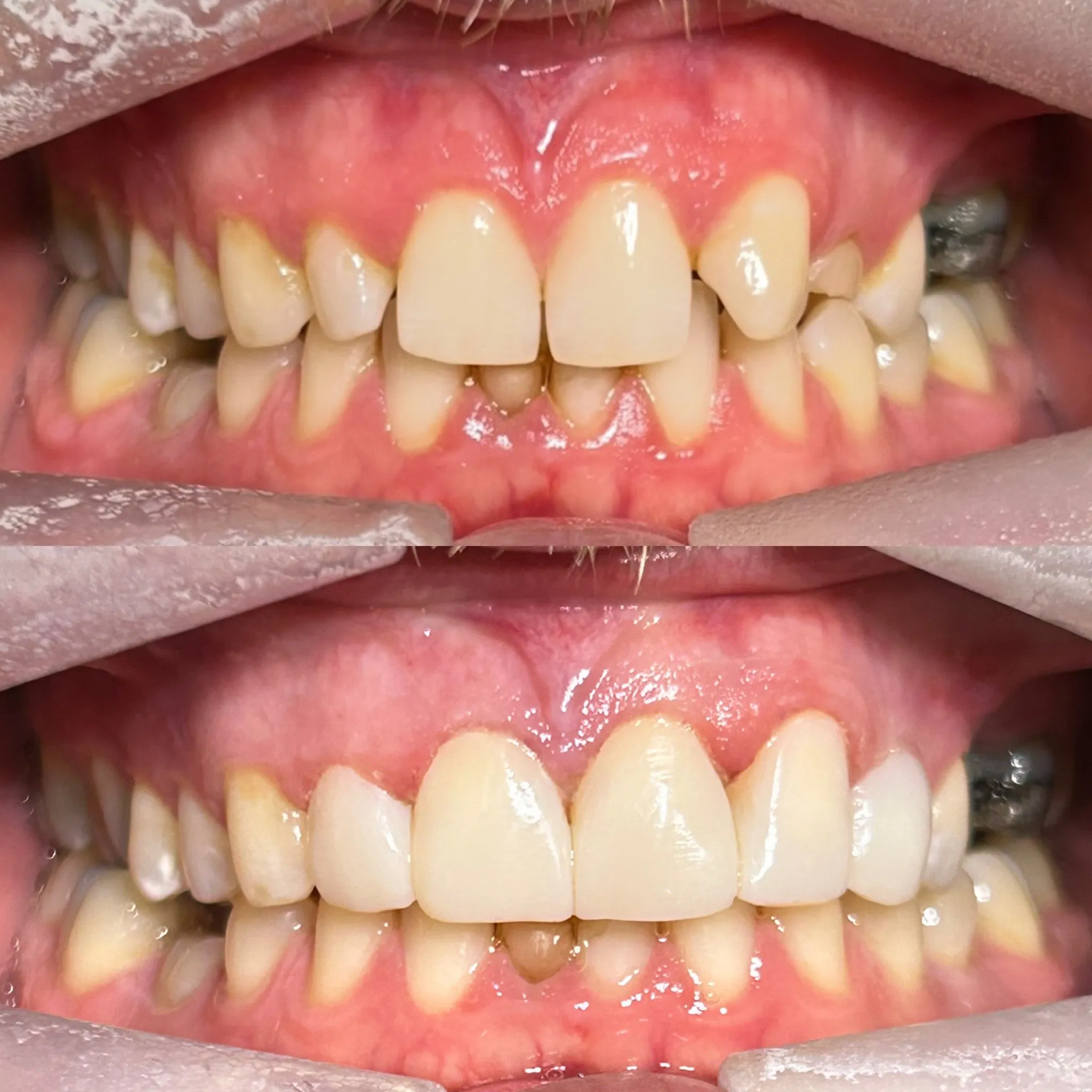
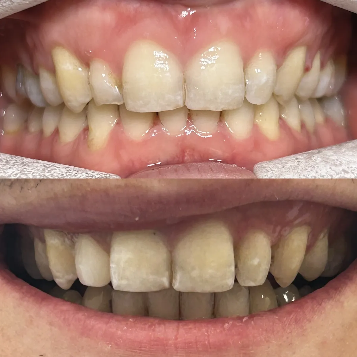
The patient attended for an adult dental examination with concerns relating to existing composite bonding on the upper anterior teeth. The medical history was reviewed and updated at today’s visit with no concerns identified. The patient is a non-smoker, reports no alcohol intake, and is assessed as low risk for oral cancer.
The patient’s chief complaints were that the existing composite bondings are chipped, rough, and misshapen, with associated bleeding at the gum margins, particularly affecting the upper anterior teeth (canine to canine).
Clinical discussion highlighted important occlusal limitations. It was explained that tooth lengthening is not suitable in this case due to the patient’s bite, as this would risk damage to restorations. Additionally, it was discussed that the lateral incisors cannot be built to the same shape or length as the central incisors, as occlusal forces would likely cause failure of the composite. However, it was explained that additional contour and body could be added to improve aesthetics while respecting the bite.
Temporomandibular joint assessment revealed clicking on opening, which was noted and taken into consideration during treatment planning.
A comprehensive staged treatment plan was outlined and agreed:
It was also discussed that a night guard (NG) would be required at a later stage to protect the restorations and manage occlusal forces.
The patient demonstrated understanding of the findings, limitations, and proposed treatment stages and is happy to proceed with the outlined plan.
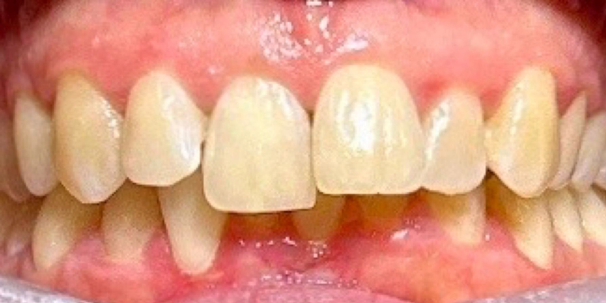
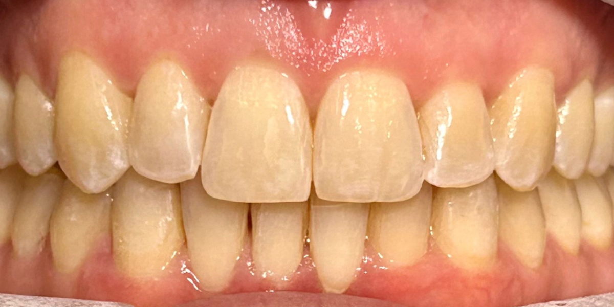
The patient presented with concerns regarding the alignment of her teeth, noting that they are not straight. She has a history of orthodontic treatment during her teenage years; however, she did not maintain retainer wear following treatment. As a child, the patient had a thumb-sucking habit, which may have contributed to her current malocclusion.
On examination, the patient exhibits a deep bite with a skeletal Class II anteroposterior relationship. Facial symmetry is within normal limits, with an average Frankfort mandibular plane angle (FMPA) and average oral hygiene. The patient reports no current oral habits.
The incisor relationship is Class II, Division 1, with the upper right central incisor (UR1) and upper left lateral incisor (UL2) demonstrating Class II positioning. The overjet is increased, measuring 7 mm at UL1 and 5 mm at UR1, and the overbite is increased and complete. Dental midlines reveal an upper midline that is centred, while the lower midline is deviated 2 mm to the right.
Molar and canine relationships are asymmetric. On the right side, both molar and canine relationships are Class II half-unit, while on the left side, molar and canine relationships are Class I. No crossbite is present.
The patient demonstrates moderate crowding in both the upper and lower arches, with no spacing noted in either arch.
Treatment options were discussed in detail. The patient is a candidate for either Invisalign or fixed orthodontic appliances. It was explained that Invisalign treatment would likely require the use of attachments, including vertical attachments, as well as interproximal reduction (IPR). A comprehensive course of treatment was recommended, and the patient was advised that Class II elastics may form part of the treatment plan. It was also discussed that there may be limitations in achieving full overjet correction with aligner therapy alone. Fixed appliances remain an alternative option should the patient prefer or require them.
An iTero digital scan was taken at today’s visit to facilitate treatment planning.
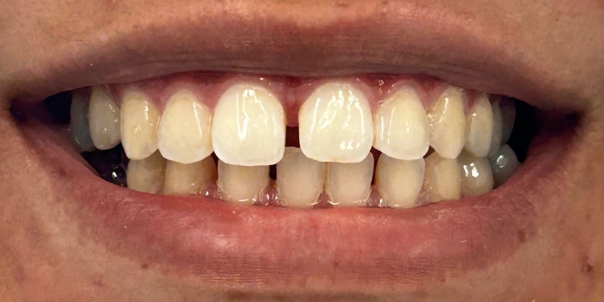
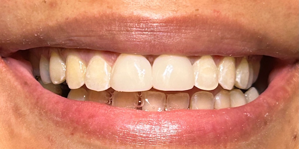
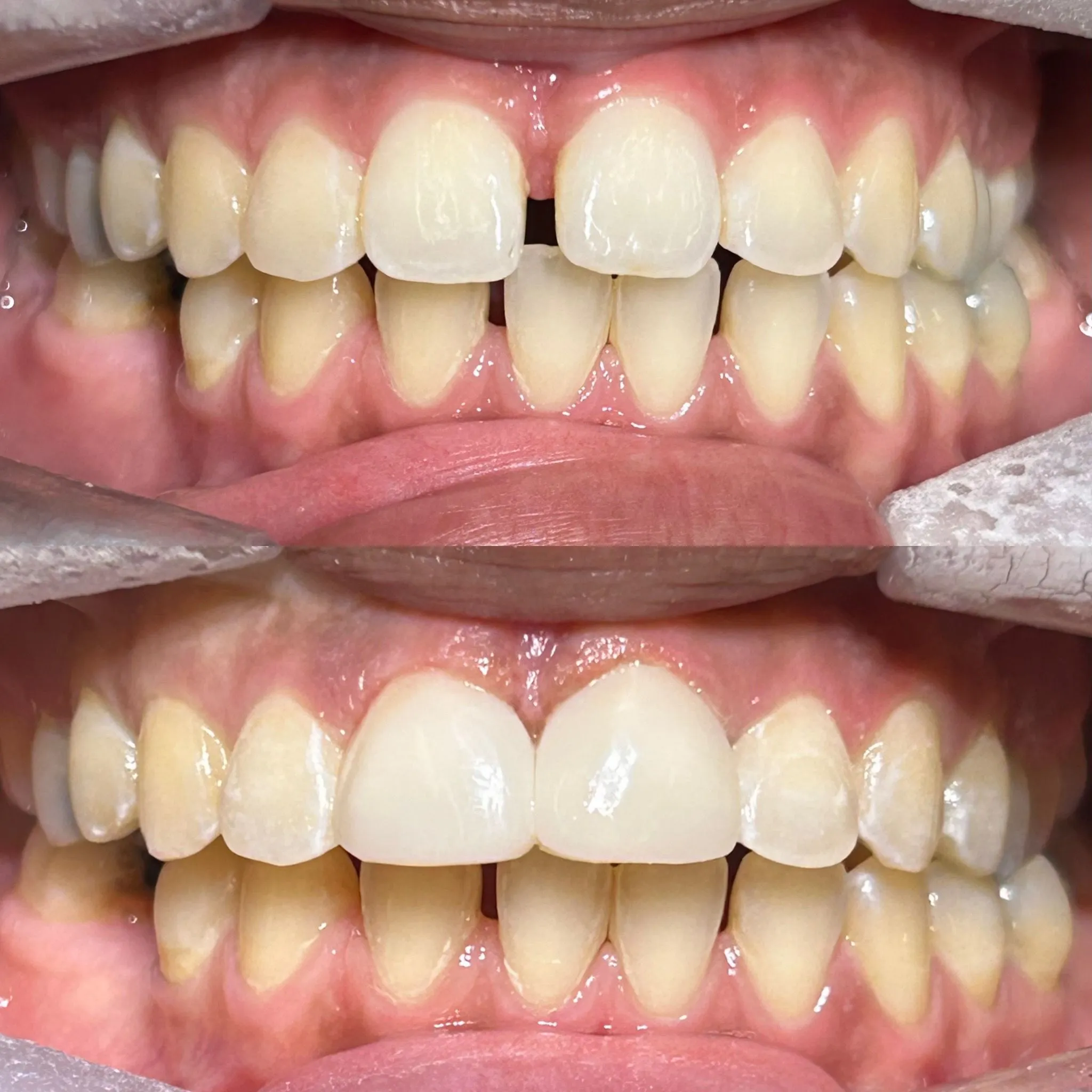
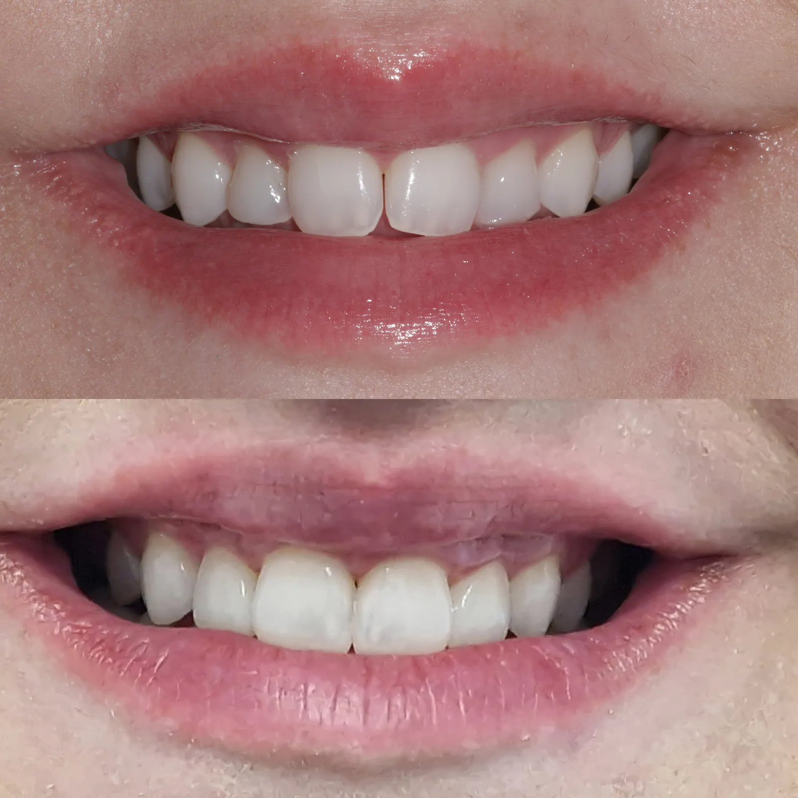
The patient presented with the chief complaint of wanting to close the gap between the upper front teeth and expressed a particular interest in composite bonding as a treatment option. The patient has her own general dentist and reported a history of orthodontic treatment on two previous occasions. Unfortunately, retainer compliance was compromised as the retainers were damaged after being chewed by her dog. The patient is not concerned about the presence of spacing in the lower arch.
The patient’s medical history is recorded separately (see medical history) and was reviewed at the appointment.
On clinical examination, it was explained that the ideal first-line treatment option to address the spacing would be orthodontic treatment. However, it was also discussed that a camouflage approach using composite bonding could be successfully achieved to close the upper anterior spaces. The limitations and benefits of this approach were explained in detail.
The patient was advised that placement of a bonded retainer would be recommended following composite bonding to support occlusal stability and help maintain the result long term.
A shade assessment was completed at today’s visit, with VITA shade B1 selected. The patient expressed interest in finance options, which were discussed and provided.The patient demonstrated understanding of the proposed treatment and will consider the options discussed.
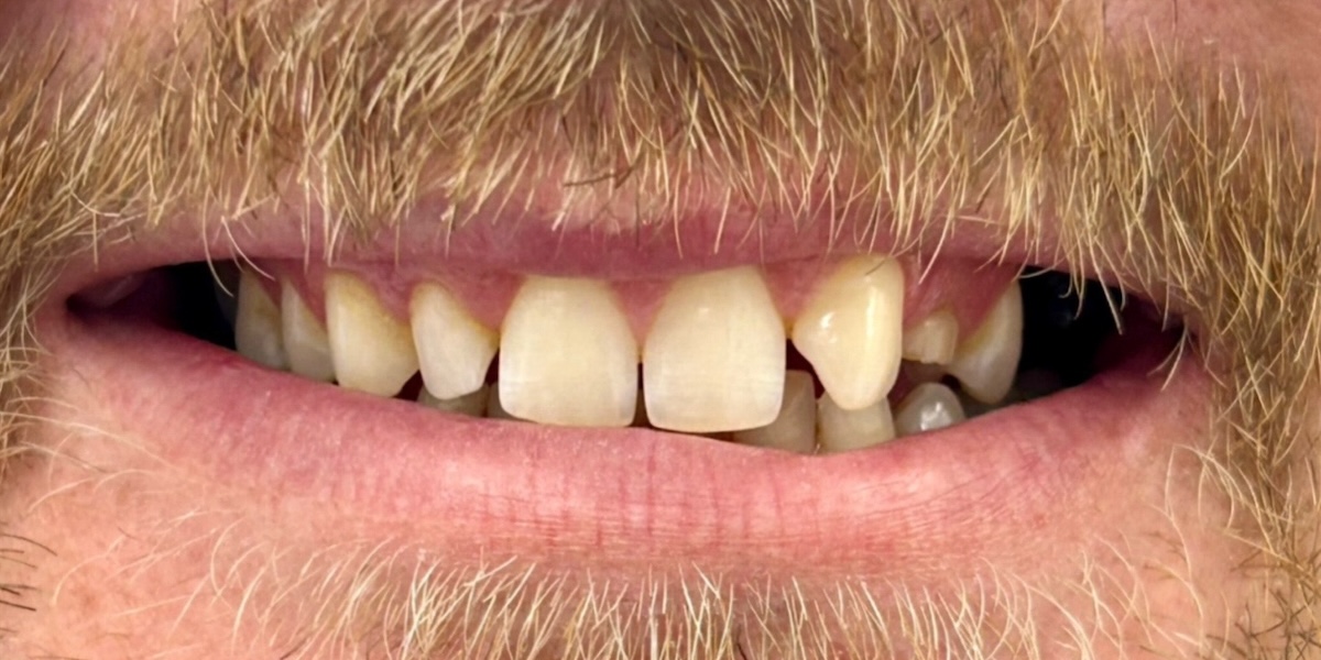
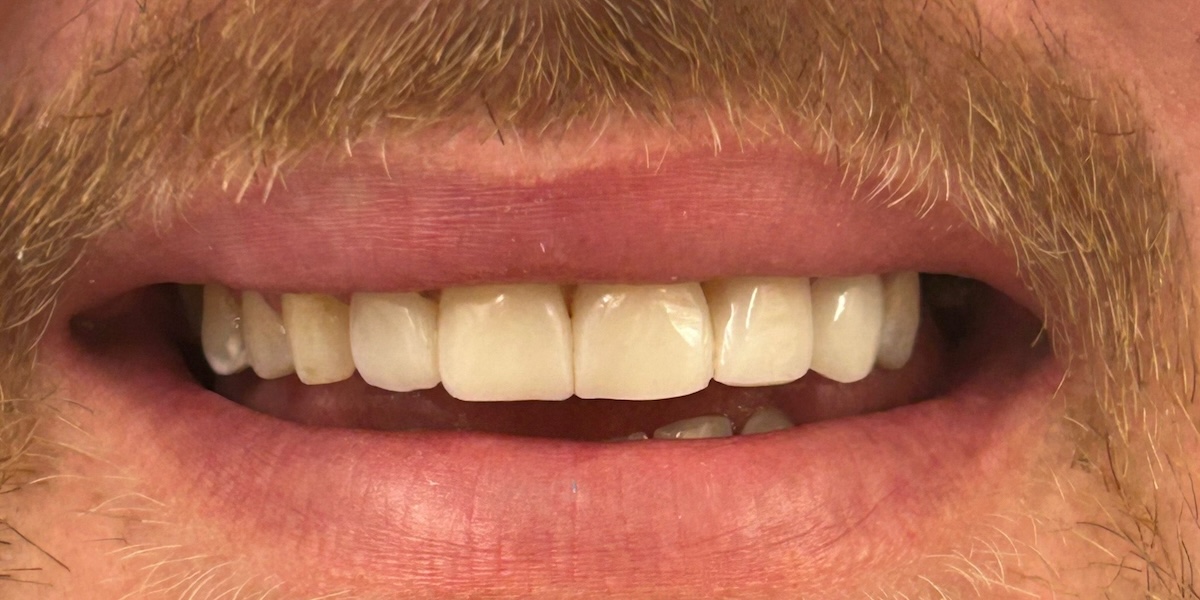
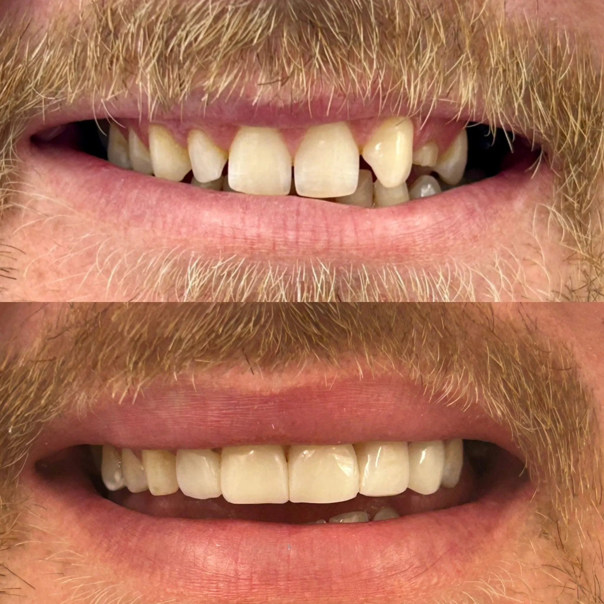
The patient attended for an examination with interest in cosmetic improvement of the upper anterior teeth.
Clinical examination revealed hypodontic enamel with mis-shapen anterior teeth. The upper left second premolar (UL5) is impacted. The occlusion demonstrates a Class I molar relationship, with canines in a Class II half-unit relationship and Class II Division 2 upper central incisors. The upper right lateral incisor (UR2) presents as a peg lateral, suitable for reshaping with composite bonding.
Soft tissue examination was within normal limits, with good moisture, no mucosal lesions, a mobile tongue, and tonsils appearing normal. Extra-oral examination was unremarkable. Temporomandibular joint assessment showed a symmetrical path of closure with full opening, although the right masseter muscle appeared enlarged. Lymph nodes and salivary glands were non-palpable and within normal limits.
Oral hygiene assessment revealed the presence of tartar and staining, and oral hygiene instruction was provided. Periodontal screening was recorded as per BPE, with an overall low periodontal risk. No tooth surface loss was noted. Bitewing radiographs are scheduled on a two-yearly basis; two bitewings were taken today (left and right) to assess caries and bone levels, both graded A.
A periapical radiograph of the UL3–UL5 region was taken, revealing an impacted UL5 in close proximity to UL4. A radiolucency on the distal aspect of the UL4 root was noted, suggestive of possible external root resorption. Clinically, UL4 did not respond to cold testing (Endo Cold). These findings were discussed with the patient, and it was explained that the prognosis of UL4 is poor, particularly in relation to future bridge work in this area.
Treatment options were discussed in detail. The patient was advised that the shape of the upper left canine (ULC) could be camouflaged with composite bonding to improve aesthetics. It was also explained that, should ULC and UL4 be lost in the future, alternative restorative options such as implant treatment could be considered. An orthodontic assessment with CBCT imaging was recommended to further evaluate the extent of resorption and potential exposure of the UL4 root; however, the patient stated that he is not interested in orthodontic treatment at this stage.
Proposed Treatment Plan:
The patient is happy to proceed with composite bonding to:
The patient demonstrated understanding of the findings, risks, benefits, and limitations discussed and wishes to proceed with the proposed cosmetic treatment.
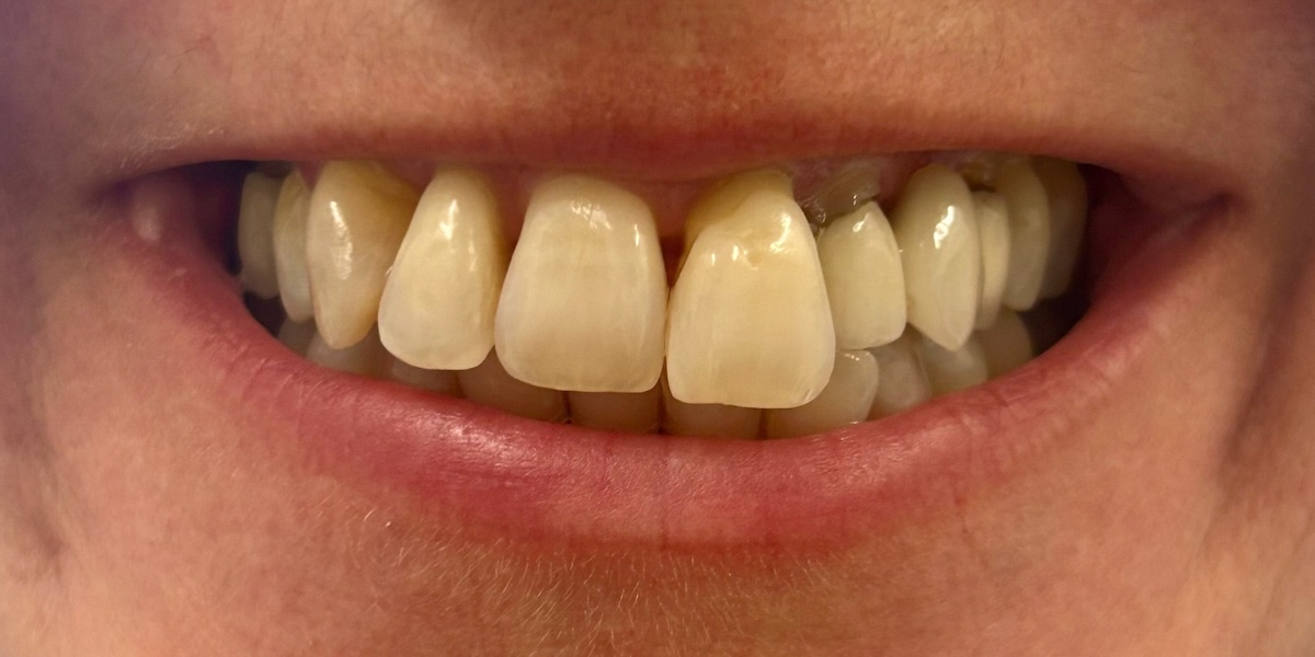
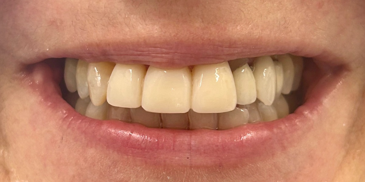
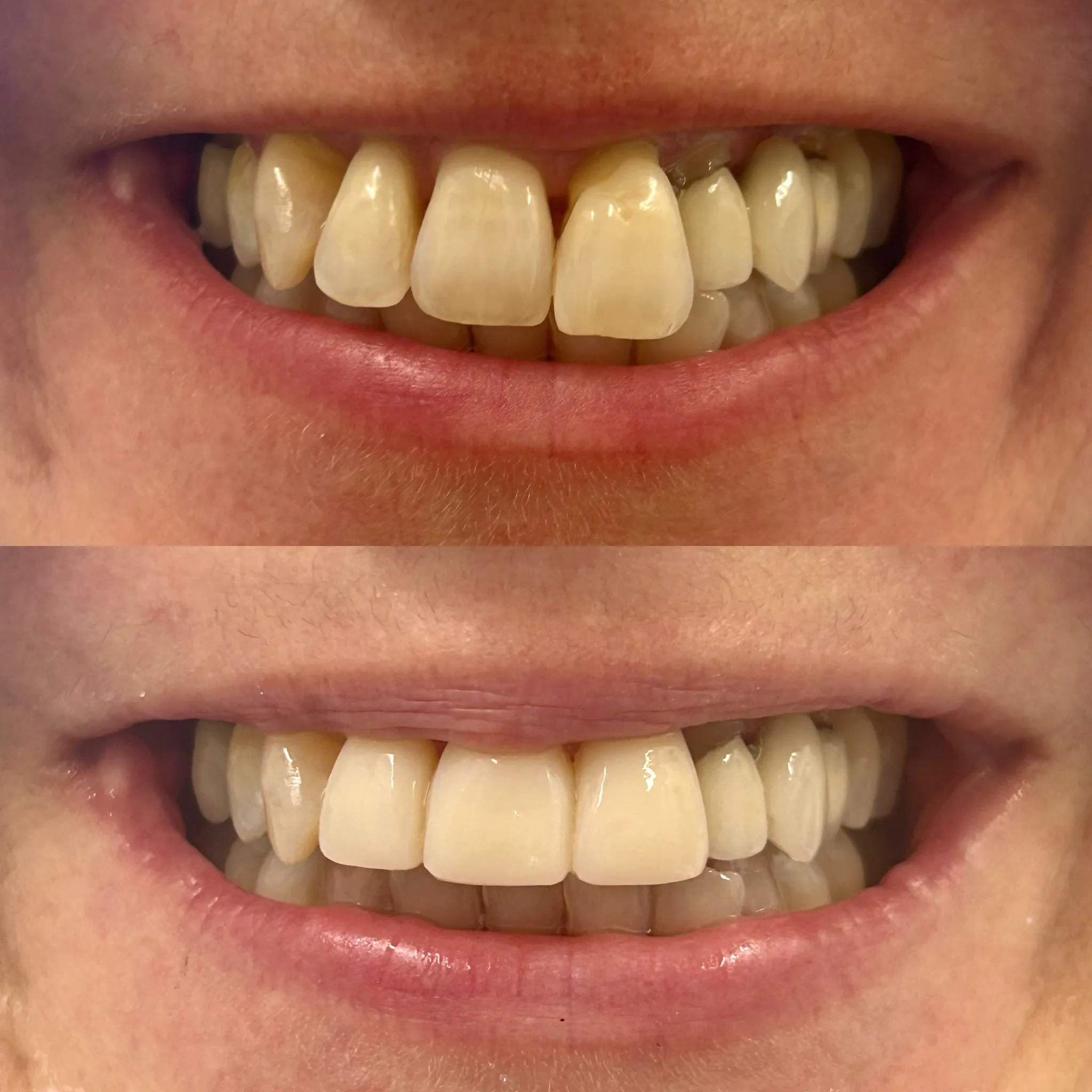
The patient attended for a composite bonding consultation and provided informed consent for the appointment. Her chief concerns relate to the upper left central incisor (UL1), which she feels is rotated and associated with localized gingival recession. She also expressed dissatisfaction with the dark margin (“black line”) along the gingival margin of the existing bridge spanning UL2–UL4. The patient stated a preference to avoid traditional silver orthodontic brackets.
On extra- and intra-oral examination, the patient demonstrates a Class I incisal and canine relationship. There is evidence of wear facets on UR1, UL1, and the UL2–UL4 bridge. The upper left lateral incisor (UL2) is noted to be extruded, though it is not mobile. No gingival recession is present in the lower arch. The patient has a high lip line on smiling, making the aesthetic outcome of treatment particularly important.
Treatment options were discussed in detail. It was explained that orthodontic treatment would be the ideal option to correct tooth position prior to any restorative work; however, composite bonding could be considered as a camouflage approach, with associated limitations discussed. The importance of retainer wear following either orthodontic or composite bonding treatment was emphasised to reduce the risk of relapse and to protect the restorations.
Clear aligner therapy was discussed as an orthodontic option, consisting of approximately 20 Invisalign aligners worn for 22 hours per day, removed only for eating. The patient was informed about upcoming open days, which may include promotional offers such as a complimentary OPG, retainers, and tooth whitening following Invisalign treatment.
The condition of the existing bridge was also discussed, and future bridge-related treatment options were outlined where relevant.
The patient demonstrated understanding of the options, risks, benefits, and limitations discussed and will consider how she wishes to proceed with treatment.

.avif)







.webp)